
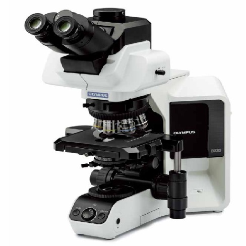
Cuihe provides enterprises with BX53 microscope, BX53 microscope price, and BX53 microscope parameter related information.
1.
Core technical features · High-fidelity color restoration (accurate without color cast): Equipped with a high-color-rendering LED light source, whose spectral characteristics are very close to natural sunlight.
Combined with Olympus high-performance objective lenses, it can achieve extremely high chromatic aberration correction, ensuring that specimens (such as stained pathological sections) observed at different magnifications have true colors and no color cast, meeting demanding clinical diagnostic needs. · Highly modular design (can be freely upgraded): It adopts a building block body structure and has strong application expandability.
Users can freely configure or add different observation modules (such as bright field, dark field, phase contrast, polarization, differential interference and multi-color fluorescence observation, multiple people can observe together) according to the actual business or scientific research direction, without changing the microscope host. · Humanized and energy-saving design: The body is equipped with an ECO infrared sensor.
When it detects that the operator has left for about 30 minutes, the system will automatically turn off the light source to extend the life of the equipment and save energy.
The fuselage structure is ergonomically designed, which greatly relieves the fatigue of long-term continuous work. · Intelligent light intensity management (no need for frequent dimming): Built-in intelligent light intensity management (LIM) function.
When the user switches objective lenses with different magnifications, the system will automatically memorize and adjust the optimal brightness corresponding to the objective lens to achieve "switching magnification and constant brightness", avoiding the problem of the field of view flickering when changing lenses in traditional microscopes. (Coded nosepiece required) Upgraded fluorescence: Relying on BX53 The basic microscopy host expands the vertical fluorescence imaging light path and adds a dedicated fluorescence excitation imaging system.
It excites the fluorescence color of the sample through specific light signals, effectively weakens stray light interference and improves the clarity of weak fluorescence imaging.
It can realize multi-color specific fluorescence label observation, clearly distinguish different labeled fluorescence signals in the sample, and supports various fluorescence imaging detections such as stained sections, live cell fluorescent proteins, and in situ hybridization.
The bright field and fluorescence observation modes can be quickly switched for comparison to meet the fluorescence characterization and image acquisition needs of biological cells and pathological tissues.
Upgraded phase contrast: Based on the BX53 basic host, a phase contrast imaging light path is built to expand the phase contrast observation function.
Transparent living samples can be observed without dyeing the samples.
The subtle refractive index differences of cells and microorganisms are converted into clear light and dark images, eliminating the problem of no contrast in transparent sample observation.
The original physiological state of the sample is completely preserved.
It supports one-click switching between conventional brightfield and phase contrast observation modes.
It is suitable for in vivo monitoring of cell culture and observation of living microbial morphology.
Upgraded DIC: Based on the BX53 main unit, it is equipped with a differential interference imaging system and upgraded differential interference observation mode.
It relies on polarized light interference to present images with a three-dimensional relief texture without halo artifacts, greatly improving the layering and resolution of microstructure.
It can clearly distinguish ultra-thin sections and submicroscopic fine three-dimensional structures of cells.
It can take into account the observation of stained and unstained samples to meet the needs of fine analysis of various high-resolution microscopic morphologies. 2.
Upgrade multi-person observation: Relying on the BX53 basic microscope host to expand the multi-person synchronous observation light path and expand the multi-person observation function, it can realize the simultaneous real-time observation of the same microscope field by multiple people.
The field of view is synchronized without deviation and does not interfere with the main observer's operation.
It can be flexibly adapted to teaching, case consultation, team discussion and other scenarios, and can be compatible with various imaging modes such as fluorescence, phase contrast, and DIC. 2.
Core application areas Medical and clinical testing: hospital pathology department (slice screening, cancer diagnosis), laboratory department (blood cell and microbial analysis), cytology examination (cervical TCT screening), assisted reproduction (ART, artificial insemination research) Biology and life sciences: cell biology research, immunofluorescence observation, neuroscience and gene chip testing in universities and scientific research institutes Industry and materials science: after configuration adjustment ( BX53M/P series), it can be used for material metallographic analysis, mineral crystal polarization identification, semiconductor defect detection, etc. (According to the material characteristics, it can be customized with special long working distance or polarizing objectives or stages, etc.) 3.
Selection of objective lenses: The imaging quality of BX53 depends on the grade of the objective lens it is equipped with.
Olympus has the industry's top optical processing technology, and its objective lenses are mainly divided into three mainstream levels.
Users can flexibly configure them according to budget and scientific research needs: Plan Achromat objective lens series (Plan Achromat): basic and practical, mainly correcting red and blue chromatic aberration.
It is cost-effective and can fully meet the needs of routine teaching, hospital clinical testing and ordinary bright field observation.
Plan semi-apochromatic (Fluar series): advanced scientific research type, using special fluorspar glass, with higher luminous flux and chromatic aberration correction capabilities.
Excellent performance in immunofluorescence imaging, capturing weak fluorescence signals against dark backgrounds.
Plan apochromatic objective lens (Apo series): top flagship model, which performs perfect chromatic aberration and spherical aberration correction for red, green, blue and other wavelengths.
BX53 is a flagship upright biological microscope designed for clinical pathological diagnosis, advanced cytological examination and cutting-edge academic research.
This model is specially designed for molecular diagnosis of tumor slices, morphological analysis of bone marrow cells and fluorescent multi-color labeling imaging of scientific research groups in the pathology department of large tertiary hospitals;
With the intelligent light intensity management (LIM) system and UIS2 advanced optical system, it can not only provide pathological images with extremely high color reproduction and resolution, but also support the expansion of multi-functional applications such as bright field, fluorescence, phase contrast, and polarization.
The automated electric stage and fully automatic scanning control software produced by our company can also be modified or customized according to actual needs to provide an excellent hardware base for the AI-assisted diagnosis system (secondary development)
BX53 is a flagship upright optical microscope positioned for clinical medicine and advanced scientific research.
As a mainstream benchmark model in the industry, it is known for its high-fidelity image quality, powerful modular expansion capabilities, and efficient intelligent operation.
It is currently the most commonly used mid-to-high-end microscope in major hospitals, universities, and scientific research institutes at home and abroad.